Back Muscles Diagram / Muscles Of The Back Teachmeanatomy : Build wide lats with this back building exercise.. Anatomynote.com found anatomy of back muscles diagram from plenty of anatomical pictures on the internet. Our latest youtube film is ready to run. The superficial and intermediate muscles do not develop in the back, and are classified as extrinsic muscles. Related posts of muscles of the lower back and buttocks diagram piriformis muscle anatomy ultrasound. The sections below cover these in more detail.
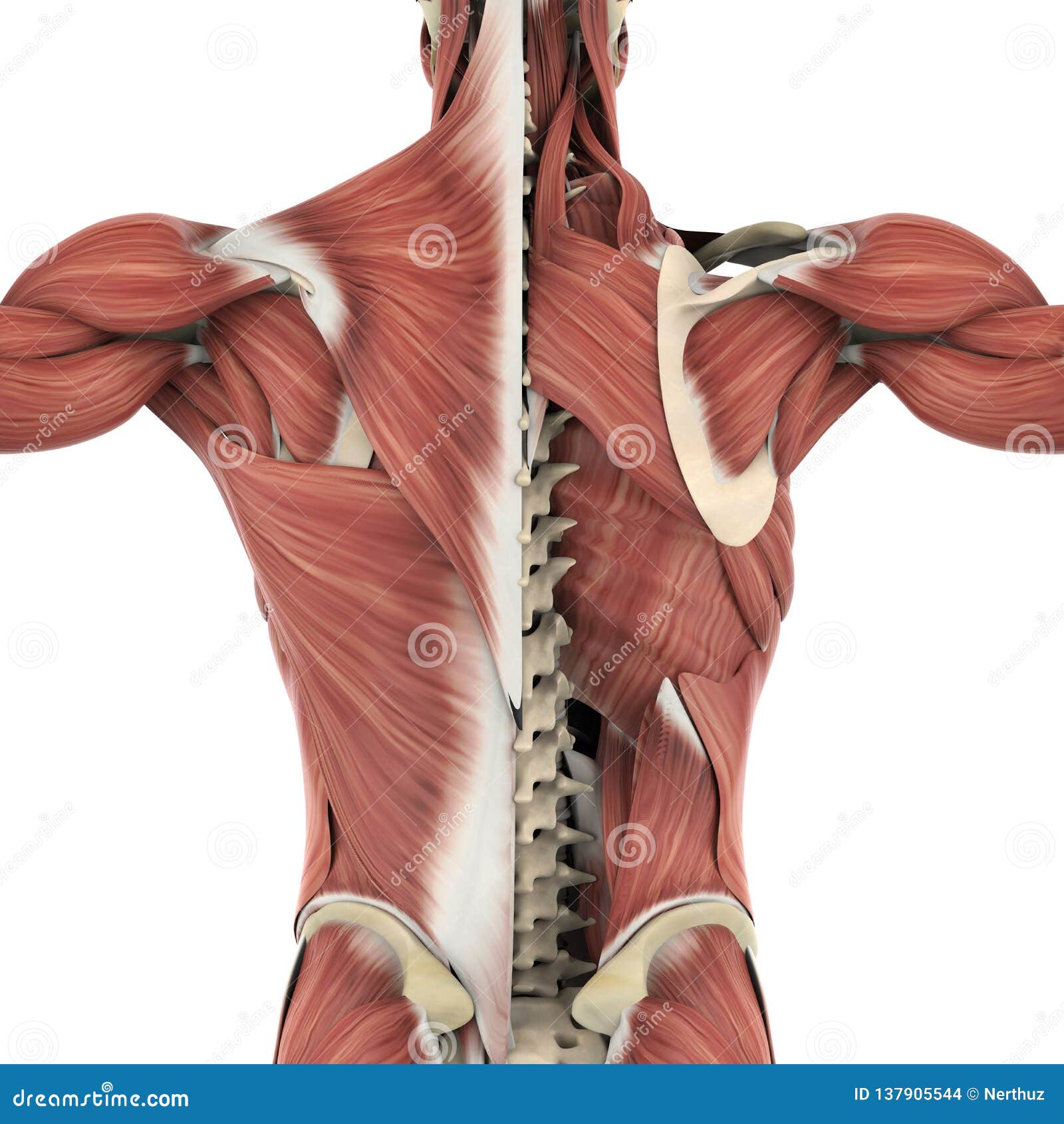
The muscles of the back with the surface (trapezius, latissimus dorsi, thoracolumbar fascia, deltoid) and intermediate layers (serrated muscles, external and internal oblique muscle). Pain log more pain mapping tools Five pairs of lumbar spinal nerves labeled l1 to l5 branch off your spinal cord and exit through small holes between the vertebrae. To learn more about the anatomy of the spine, watch this video. The superficial and intermediate muscles do not develop in the back, and are classified as extrinsic muscles.
Muscles Of The Back Teachmeanatomy from teachmeanatomy.info The superficial and intermediate muscles do not develop in the back, and are classified as extrinsic muscles. They are in fact different, but all three work together to support your spine and to help protect it from injury. It is opposite from the chest, and the vertebral column runs down the back. Chronic back pain map this tool recommended for: Five pairs of lumbar spinal nerves labeled l1 to l5 branch off your spinal cord and exit through small holes between the vertebrae. People with back pain people who experience headaches printing for use during doctor visits to communicate information about your symptoms quickly tracking your progress over time related tools: Human muscle system, the muscles of the human body that work the skeletal system, that are under voluntary control, and that are concerned with movement, posture, and. The sections below cover these in more detail.
The superficial and intermediate muscles do not develop in the back, and are classified as extrinsic muscles.
Nerves in your lower back. Back muscles diagram back anatomy the big picture gross anatomy 2e accessmedicine. Both the deltoid and the trapezius are firmly attached to the spine of the scapula. The back anatomy includes the latissimus dorsi, trapezius, erector spinae, rhomboid, and the teres major. Postural and active movement muscle, used to tilt and turn the head and neck, shrug, steady the shoulders, and twist the arms. Extrinsic and intrinsic.the back functions are many, such as to house and protect the spinal cord, hold the body and head upright, and adjust the movements of the upper and lower limbs. Lower back muscle diagram anatomy does degenerative disc disease affect the lower back muscle? It is opposite from the chest, and the vertebral column runs down the back. The sections below cover these in more detail. The back has a total of 40 muscles. The muscles of the back with the surface (trapezius, latissimus dorsi, thoracolumbar fascia, deltoid) and intermediate layers (serrated muscles, external and internal oblique muscle). Back muscle diagram back muscles big back big back muscles big lats bodybuilding secrets major back muscles. For more anatomy content please follow us and visit our website:
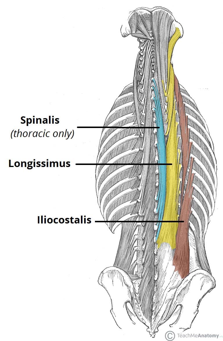
Superficial, intermediate, deep and deepest layers.these muscles lie on each side of the vertebral column, deep to the thoracolumbar fascia they span the entire length of the vertebral column, extending from the cranium to the pelvis Female reproductive organs front view. Most of the time, back muscle pain is diagnosed then treated with little more than a prescription of rest, painkillers and muscle relaxants. Human anatomy · may 25, 2021. Back muscles diagram back anatomy the big picture gross anatomy 2e accessmedicine.
Anatomy Back Muscles Stock Illustrations 1 405 Anatomy Back Muscles Stock Illustrations Vectors Clipart Dreamstime from thumbs.dreamstime.com The superficial and intermediate muscles do not develop in the back, and are classified as extrinsic muscles. The muscle elevates, depresses, rotates, and retracts the scapula, or shoulder blade. Human anatomy · may 25, 2021. There are three different groups of muscles in the back. Back to tracking tools main page. Related posts of muscles of the lower back and buttocks diagram piriformis muscle anatomy ultrasound. Postural and active movement muscle, used to tilt and turn the head and neck, shrug, steady the shoulders, and twist the arms. Build wide lats with this back building exercise.
Just need a glimpse, leave your valuable advice let us know , and subscribe us!
This muscle is a major generator of lower back and hip pain, as well as being responsible for complaints of a burning sensation along the posterior superior iliac spine (psis) and sacroiliac joint. It is opposite from the chest, and the vertebral column runs down the back. The deep muscles develop embryologically in the back, and are thus described as intrinsic muscles. The back is the body region between the neck and the gluteal regions. These muscles include the large paired muscles in the lower back, called erector spinae, which help hold up the spine, and gluteal muscles. Others, like sumo deadlifts, have been shown in emg studies—and in the trenches—to focus more on other muscle groups than the back. Build wide lats with this back building exercise. The superficial and intermediate muscles do not develop in the back, and are classified as extrinsic muscles. Chronic back pain map this tool recommended for: By the way, have you heard about the myth of. The deep back muscles, also called intrinsic or true back muscles, consist of four layers of muscles: The muscles of the back can be arranged into 3 categories based on their location: Another common cause of lower back and hip pain is disc injury.
Five pairs of lumbar spinal nerves labeled l1 to l5 branch off your spinal cord and exit through small holes between the vertebrae. We are pleased to provide you with the picture named muscles of lower back diagram.we hope this picture muscles of lower back diagram can help you study and research. We think this is the most useful anatomy picture that you need. For more anatomy content please follow us and visit our website: This is a diagram of the larger and more surface muscles of the low back.
Labeled Anatomy Chart Of Neck And Back Muscles On White Background Stock Photo Download Image Now Istock from media.istockphoto.com Pain log more pain mapping tools For more anatomy content please follow us and visit our website: The back is the body region between the neck and the gluteal regions. Back to tracking tools main page. Both the deltoid and the trapezius are firmly attached to the spine of the scapula. Likewise, there are muscles in other parts of the body that help support and move the spine. Anatomynote.com found anatomy of back muscles diagram from plenty of anatomical pictures on the internet. The sections below cover these in more detail.
Human anatomy for muscle, reproductive, and skeleton.
Female reproductive organs front view. Just need a glimpse, leave your valuable advice let us know , and subscribe us! The extensor muscles are attached to back of the spine and enable standing and lifting objects. People with back pain people who experience headaches printing for use during doctor visits to communicate information about your symptoms quickly tracking your progress over time related tools: Facebook twitter google+ linkedin stumbleupon tumblr pinterest reddit vkontakte share via email print. The muscles of the back with the surface (trapezius, latissimus dorsi, thoracolumbar fascia, deltoid) and intermediate layers (serrated muscles, external and internal oblique muscle). This is a diagram of the larger and more surface muscles of the low back. For more anatomy content please follow us and visit our website: How many muscles are in the back? Anatomynote.com found anatomy of back muscles diagram from plenty of anatomical pictures on the internet. The back has a total of 40 muscles. Below you'll see diagrams along with the names of the back muscles that may be the cause of your pain. They are in fact different, but all three work together to support your spine and to help protect it from injury.